At our centre, we use the most modern neurosurgical operating techniques to reach and remove the tumour with maximum protection of healthy brain tissue. This includes the following established techniques and aids:
COVID-19 –> information and vaccination centre
The primary goal of surgical treatment of brain tumours is to remove the tumour as completely as possible without damaging healthy tissue.
At our centre, we use the most modern neurosurgical operating techniques to reach and remove the tumour with maximum protection of healthy brain tissue. This includes the following established techniques and aids:
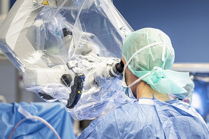
All operations on the brain are performed under a state-of-the-art surgical microscope. This enables even the smallest structures such as blood vessels to be recognised.
The use of an intraoperative navigation system can facilitate or supplement the planning of the access route to the tumour and the spatial orientation during the operation. Here, the tissue part to be treated can be visualised and controlled with previously prepared sectional images (MRI / CT / PET) even before it is freely visible.
Rapidly and aggressively growing brain tumours (gliomas) have no sharp border with healthy brain tissue. In order to achieve the most radical possible removal of the tumour tissue, the rapidly dividing tumour cells are marked with a special dye based on the amino acid 5-ALA before the operation. The dyed tumour areas can then be clearly displayed and removed.
For a few tumour operations - especially those close to the brain regions responsible for speech formation and understanding speech - it may be useful to wake the patient up during the operation and perform speech tasks with him/her. The aim is to achieve the greatest possible removal of the tumour while preserving the speech areas. During the operation, the patient is accompanied by a team consisting of the neurosurgeon, experienced anaesthetists and speech therapists.
Endoscopes are instruments that are connected to a camera system and a light source via lens optics. This enables operations to be performed using the smallest and most gentle accesses. Common areas of application are the treatment of tumours in the area of the pituitary gland (hypophysis) and diseases within the cerebral ventricles.
Neuromonitoring (IONM) is used for continuous monitoring of some neurological functions during anaesthesia for operations on the brain and spine. This technique helps to avoid neurological complications. Through targeted stimulation, the functionality of e.g. the motor and emotional pathways as well as individual important nerves can be checked without any relevant time delay.
This is another method that can be used in selected cases to visually represent the extent and boundaries of brain tumour tissue.
In the few cases where tumour resection is not possible or necessary, we perform a targeted navigation-assisted needle biopsy.