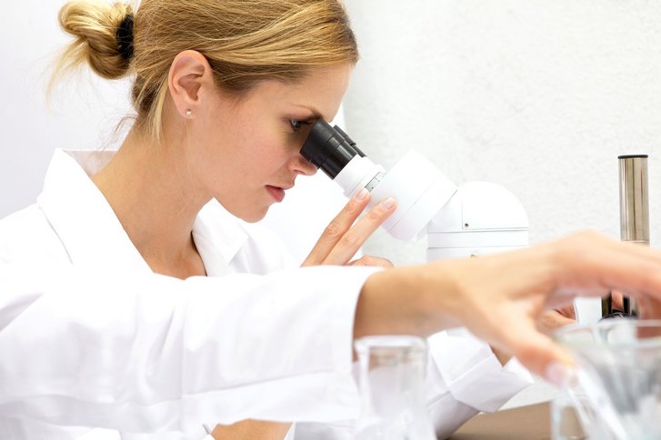
The microscopic evaluation of tissue samples of the skin and subcutaneous tissue (dermatohistopathology) is a main focus of our clinic. In the case of inflammatory skin diseases, we use microscopic examination in order to be able to more reliably assess unclear diagnoses from the direct synopsis of the clinical and histological image. Special staining as well as immunohistology or immunofluorescence help to find a diagnosis, especially in difficult cases.
Fine tissue examination for skin tumours
A further field of application of dermatohistopathology is the diagnostic (3-D histology) of skin tumors. The fine tissue examination provides essential information on the type and classification of tissue changes and is of decisive importance for the further surgical or therapeutic procedure, especially in the case of malignant tumours.
With the help of the so-called rapid embedding, we can assess on the day after the surgical removal whether a skin tumour has been sufficiently removed in a healthy person or whether a follow-up operation is still necessary. Above all, our patients at the Skin Cancer Center benefit from this particularly fast and highly specialised car
