COVID-19 –> information and vaccination centre

Imaging of the heart: Cardio-MRI and Cardio-CT

Imaging of the heart: Cardio-MRI and Cardio-CT

We offer our patients non-invasive diagnostics by means of cardio-MRI and cardio-CT. Both procedures enable a highly specialised examination of the heart.

Cardio-MRI Centre

With cardio-magnetic resonance imaging (abbreviated Cardio-MRI), we offer you one of the most modern non-invasive examination procedures in cardiology. It is a low-risk procedure for the patient without radiation exposure and allows us to make precise statements about the anatomy and functioning of the heart.

As Cardio-MRI Centre, we perform about 2,000 examinations a year. We use a radiological device with special equipment for cardiac and vascular diagnostics. The entire examination takes 20-45 minutes, depending on the problem, and can easily be performed on an outpatient basis.

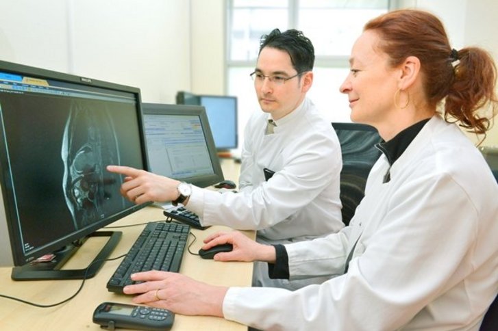
In Cardio-MRI, radiologists and cardiologists work hand in hand. On the cardiological side, Dr. Brigitte Bathgate takes care of our patients. She has the highest certification in the field of cardiac MRI according to the European Society of Cardiologists (ESC) and has extensive experience in the diagnostic evaluation of the sectional images.

  • Left ventricular and atrial parameters
  • Coronary artery disease
  • Circulatory disorders of the coronary vessels
  • Differentiation of acute from chronic myocardial infarction
  • Extension of scars
  • All forms of cardiomyopathies (non-inflammatory myocardial diseases)
  • Inflammatory myocardial diseases
  • Cardiac involvement of systemic diseases
  • Pericardial diseases
  • Valve diseases (stenoses and insufficiencies)
  • Congenital heart disease in children and adults
  • Cardiac space-occupying lesions (thrombi and tumours)
  • Vascular (coronary anomalies, aortic diseases and pulmonary veins)


Cardio-CT: Computed tomography of the heart

The latest generation of low-radiation CT scanners allows complete imaging of the heart and coronary arteries in just a few seconds in order to reliably rule out possible constrictions or occlusions. The X-ray dose is very low. The device is especially suitable for imaging the heart and the vessels close to the heart and also enables the imaging and measurement of coronary calcifications. 

The examination is carried out jointly by radiologists and cardiologists, the image data obtained is extrapolated into three-dimensional image data sets and then evaluated. The findings are followed by an individual therapy recommendation for each patient. 

During the examination you lie relaxed on your back, your head is not in the tube. You will be professionally supervised and can contact us at any time. While the actual taking of the pictures only takes a few seconds, you need to plan about 20 minutes for the entire examination process.

  • Suspicion of coronary artery disease with low to moderate clinical probability
  • Follow-up of coronary artery bypass grafting
  • Coronary abnormalities


International Services

Telefon

+49 2151 32-10845 | info@helios-international.com

Öffnungszeiten

Monday - Thursday 8:00 am to 5:00 pm
Friday 8:00 am to 2:30 pm